Describe the Process of a Simple Stain Using Methylene Blue
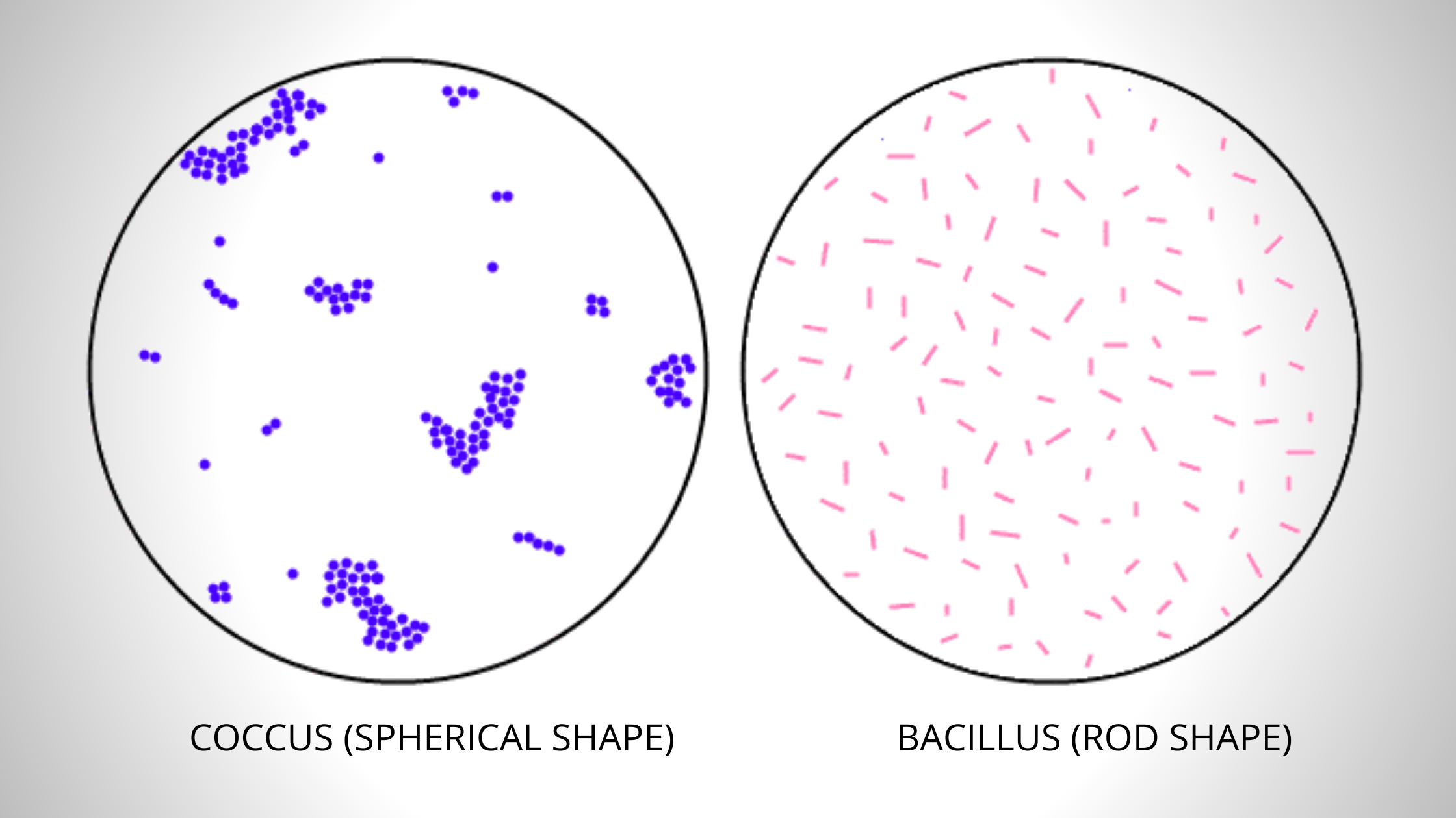
The simple stain can be used as a quick and easy way to determine cell shape size and arrangements of bacteria. Composition of Methylene Blue Stain.
What Is Simple Staining Definition Principle Procedure Video Biology Reader
If the cell is dead there will be no reaction as the cells enzymes have been inactivated.

. In a negative staining technique a negatively charged stain colors the background. Microscopic view of Bacillus rod shaped bacteria simple stained with crystal violet. Dissolve the stain in about 30 ml of water.
Any basic dye such as methylene blue safranin or crystal violet can be used to color the bacterial cells. Hold slide at 45 add decolorizing agent drop by drop until color stops running 6. Weigh 05 gm methylene blue on a piece of clean paper pre-weighed.
Methylene blue - 1 minute Crystal violet - 30 seconds Carbol fuchsin - 20 seconds During the staining the slide may be placed on the rack or held in the fingers. Cover the smear with methylene blue. Learn vocabulary terms and more with flashcards games and other study tools.
Shake off excess water and blot slide dry with bibulous paper. You may use crystal violet safranin or methylene blue. The first step in gram staining is to stain the slide with Methylene Blue dye.
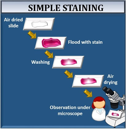
True to its name the simple stain is a very simple staining procedure involving a single solution of stain. Methylene blue 1 to 2 minutes. Flood the smear with methylene blue allow for 2 minutes pour off the stain and allow the air to dry by keeping in a slanting position and by this the organism will retain the methylene blue stain.
This is due to the cells enzymes which reduce the methylene blue causing it to lose its color. Hinfluenzae in CSF Gonococci in urethral pus Polychrome Methylene Blue. Crystal violet 20 to 60 seconds.
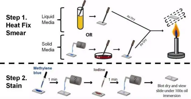
Prepare a heat fixed smear of the culture you wish to examine. Carbol fuchsin 15 to 30 seconds. Distilled water 1000 ml.
The single dye used here in our lab is methylene blue a basic stain. Saturate the smear with basic dye for approximately 1 minute. Rinse the slide gently with water.
Methylene blue staining is used to make out clearly the morphology of the organisms eg. Procedure of Simple Staining. Gently wash the smear with tap water to remove excess stain.
Stain the smear by flooding it with one of the staining solutions and allowing it to remain covered with the stain for the time designated below. After about one minute gently wash off excess dye with water. Add a basic dye usually methylene blue or crystal violet to a heat-fixed cooled bacterial smear.
Place a slide on the staining tray and flood the smear with one of the indicated stains using the appropriate exposure time for each. Perform a bacterial smear as discussed in Figure 3-52 on page 150 of your lab manual. Method of Staining Flood the smear with methylene blue allow for 2 minutes pour off the stain and allow the air to dry by keeping in a slanting position and by this the organism will retain the methylene blue stain Use Methylene blue staining is used to make out clearly the morphology of the organisms eg.
How to stain with methylene blue. Isolated and imaged by Muntasir Alam University of Dhaka. Simple Stain Simple stains provide a quick and easy way to determine cell shape size and arrangement.
Rinse slide with water to remove excess stain 5. Blot water from slide with bibulous paper 10. Rinse slide with water to remove excess stain 9.
Cover smear with methylene blue for 2 min 8. Gently wash the slide with distilled water drain off excess water blot do not rub with absorbent paper and allow slides to air dry completely. Simple Direct Staining with Methylene Blue.
Methylene blue is a simple stain that colors cells blue. Transfer the stain to a clean brown bottle. Basic stains having a positive charge bind strongly to negatively charged cell components such as bacterial nucleic acids and cell walls.
View the following clip. Finally add the remaining water. Leave on for 1-3 minutes.
Methylene blue 05 gm. Start studying Simple stain lab quiz. A thin film of bacteriamicrobe spread on a slide.
Hinfluenzae in CSF Gonococci in urethral pus. If methylene blue stain is applied to a sample a healthy cell with turn the stain colorless. Methylene blue blue Gram safranin pinkred Gram crystal violet purple.
Preparation of Methylene Blue Stain. Methylene blue staining is useful in determining cell mortality. What is a smear.
Review the steps used when preforming a simple stain or direct stain. After performing the bacterial smear smearing fixing Place the slide on a staining rack and flood it with methylene blue. Allow the dye to remain on the smear for approximately 1 minute.
Pass the slide film-side up through the flame of the bunsen burner 3 or 4 times to heat-fix. List at least three terms that are used to describe morphological. The following step often known as fixing the dye requires employing iodine to produce a Methylene blue-iodine combination to inhibit dye removal.
Immediately rinse slide to remove decolorizer 7.

Simple Staining Techniques In Microbiology
Simple Staining Principle Procedure Uses Microbe Online
Methylene Blue Stain Introduction Principle Composition Preparation
Simple Staining Procedure Principle Result


Comments
Post a Comment